- Menu
-
- English


Views: 0 Author: Site Editor Publish Time: 2026-08-11 Origin: Site


Establishing reliable vascular access is a non-negotiable foundation for emergency resuscitation and long-term therapeutic interventions. A vascular access procedure involves inserting a flexible, sterile plastic tube or catheter into a blood vessel. This pathway facilitates blood sampling, hemodynamic monitoring, and the delivery of medications, nutrients, and blood products directly into the circulatory system.
Poor device selection carries severe operational and clinical consequences. Choosing an inappropriate catheter leads to premature device failure, central line-associated bloodstream infections (CLABSI), irreversible vessel depletion, and extended hospital stays. These complications compromise patient safety and strain institutional resources. To navigate these challenges, clinical directors and procurement teams require a structured evaluation framework. This framework matches specific access devices to patient acuity levels, anticipated therapy durations, and strict institutional infection control standards.
Device selection must be driven by infusate characteristics (osmolarity, pH, vesicant potential) and projected duration of therapy to minimize complications.
Peripheral access is optimal for short-term, non-irritating therapies, while central access is mandatory for hemodynamic monitoring, prolonged treatments, and caustic medications.
Standardizing procurement of vascular access devices alongside broader critical care medical supplies reduces practice variation and improves compliance with insertion bundles.
Mitigating implementation risks requires continuous clinician training on ultrasound-guided insertion, rigorous site maintenance protocols, and comprehensive patient education.
Defining the baseline requirements for successful catheterization is a mandatory first step before evaluating specific devices. Clinical success relies on preserving the patient's venous anatomy while ensuring the prescribed therapy can be delivered without interruption or complication. A systematic approach prevents the common error of defaulting to the most familiar device rather than the most appropriate one. You must evaluate the patient's complete clinical picture, including their medical history, current diagnosis, and the specific pharmacological agents ordered by the attending physician.
The projected timeline of intravenous treatment heavily dictates device selection. Short-term needs, defined as under five days, generally require less invasive approaches like standard peripheral IVs. Medium-term requirements spanning 6 to 14 days introduce the need for more durable materials and deeper vessel placement, such as midline catheters, to avoid repeated insertions. Long-term therapies lasting months to years demand specialized devices designed for maximum durability, minimal infection risk, and seamless integration into the patient's daily life, such as tunneled catheters or implantable ports.
Evaluating the chemical properties of the medication or solution dictates whether a peripheral or central line is required. Clinicians must analyze the pH, osmolarity, and vesicant properties of the infusate. Solutions with extreme pH levels, high osmolarity, or known vesicant properties cause severe endothelial damage if administered through small peripheral veins. These caustic therapies strictly require central administration, where high blood flow rapidly dilutes the solution, preventing chemical phlebitis and tissue necrosis.
Infusate Characteristic | Examples | Recommended Access Route | Clinical Rationale |
|---|---|---|---|
Isotonic, Neutral pH (Non-vesicant) | Normal Saline, Lactated Ringer's, standard antibiotics | Peripheral (PIVC or Midline) | Low risk of endothelial damage; adequate hemodilution in peripheral veins. |
Hyperosmolar (>900 mOsm/L) | Total Parenteral Nutrition (TPN), 3% Hypertonic Saline | Central (PICC, CVC, Port) | Requires rapid hemodilution in the superior vena cava to prevent severe phlebitis. |
Vesicant / Irritant | Vasopressors (Norepinephrine), specific chemotherapy agents | Central (PICC, CVC, Port) | High risk of tissue necrosis and extravasation injuries if peripheral veins rupture. |
Extreme pH (<5 or >9) | Vancomycin, Phenytoin, Promethazine | Central (PICC, CVC, Port) | Causes chemical burns to the tunica intima of small peripheral vessels. |
The concept of Vessel Health and Preservation (VHP) emphasizes proactive planning to protect a patient's venous real estate. In complex or chronically ill patients, repeated peripheral punctures lead to rapid vessel depletion, scarring, and loss of future access sites. High-acuity patients require a strategic approach that minimizes needle sticks. Utilizing advanced imaging, such as point-of-care ultrasound (POCUS), and appropriate device scaling ensures the right line is placed successfully on the first attempt. Preserving veins in the upper extremities is especially important for patients who may eventually require hemodialysis fistulas.
Peripheral devices are the most common entry point into the vascular system, utilized primarily for straightforward, short-duration therapies. While ubiquitous, their deployment requires careful consideration of the patient's peripheral vein quality and the specific characteristics of the ordered infusions. Proper site selection, typically starting distally in the upper extremities and moving proximally, helps preserve venous options.
Short peripheral catheters are the standard choice for basic hydration, the administration of non-vesicant medications, and therapies with short dwell times, typically lasting less than four days. They are inserted into the superficial veins of the hands or arms. The primary advantage of PIVCs is their high ease of insertion, often performed rapidly at the bedside by nursing staff. However, they carry a high risk of mechanical phlebitis, fluid infiltration, and accidental dislodgement, necessitating frequent site monitoring and routine replacement. When placing a PIVC, clinicians should follow these steps:
Apply a tourniquet 4 to 6 inches above the intended insertion site to engorge the vein.
Palpate the vein to assess its depth, straightness, and condition (avoiding hard, sclerotic, or rolling veins).
Cleanse the site with a chlorhexidine gluconate solution using a back-and-forth friction scrub for at least 30 seconds.
Insert the catheter bevel up at a 10 to 30-degree angle, observing for a primary blood return in the flash chamber.
Lower the angle, advance the catheter slightly to ensure the tip is fully within the lumen, and then thread the catheter off the needle.
Secure the device with a sterile, transparent semi-permeable dressing and flush with normal saline to confirm patency.
Midline catheters serve as a vital bridge between short peripheral lines and central lines. They are inserted into the upper arm, with the catheter tip terminating in the axillary vein, short of the central circulation. Midlines are indicated for medium-term therapies lasting up to 29 days where central access is not clinically justified. They significantly reduce the need for repeated needle sticks in patients with difficult venous access. Despite their longer dwell time, midlines are still classified as peripheral devices and remain restricted from delivering continuous vesicants or highly hyperosmolar solutions. They are an excellent option for extended courses of compatible IV antibiotics.
Central venous access devices terminate in the lower third of the superior vena cava or the right atrium. This placement provides immediate hemodilution for irritating medications and allows for rapid, high-volume fluid resuscitation. Selecting the appropriate CVAD depends on the insertion environment, the required number of lumens, and the expected duration of use. Central lines require strict adherence to sterile techniques during insertion and maintenance to prevent life-threatening bloodstream infections.
PICCs are inserted through a peripheral vein in the upper arm (typically the basilic or cephalic vein) but advance until the tip reaches the central venous system. They are highly effective for long-term antibiotics, chemotherapy, TPN administration, and frequent blood sampling. PICCs offer a lower insertion risk compared to direct neck or chest central lines and are highly suitable for outpatient or home care settings. However, they carry a specific risk profile for upper extremity deep vein thrombosis (DVT), requiring careful assessment of the catheter-to-vein ratio prior to insertion. The catheter should occupy no more than 45% of the vein's internal diameter.
Non-tunneled CVCs are typically inserted directly into the internal jugular, subclavian, or femoral veins. They are the standard of care in acute critical care environments for rapid volume resuscitation, continuous hemodynamic monitoring (such as central venous pressure), and the simultaneous administration of multiple incompatible infusions. While they offer high flow rates and multi-lumen capabilities, they also present the highest risk of CLABSI and immediate insertion complications, such as pneumothorax or accidental arterial puncture. Femoral placement is generally avoided unless absolutely necessary due to the high risk of contamination and infection.
For intermittent, very long-term therapies, tunneled catheters and implantable ports are the preferred modalities. These devices are surgically placed. Tunneled catheters travel under the skin before entering the vein, creating a physical barrier against bacterial migration. A Dacron cuff on the catheter promotes tissue ingrowth, further securing the line and blocking pathogens. Implantable ports reside entirely beneath the skin and are accessed via a specialized non-coring needle (Huber needle). These options provide the lowest infection risk among CVADs, making them ideal for oncology patients and those dependent on long-term parenteral nutrition, though they require surgical intervention for both insertion and removal.
CVAD Type | Typical Insertion Site | Expected Dwell Time | Primary Clinical Indication |
|---|---|---|---|
PICC | Basilic or Cephalic Vein (Upper Arm) | Weeks to Months | Outpatient IV antibiotics, TPN, Chemotherapy |
Non-Tunneled CVC | Internal Jugular or Subclavian Vein | Days to Weeks | ICU resuscitation, multiple incompatible drips, CVP monitoring |
Tunneled CVC | Subclavian or Internal Jugular (tunneled on chest) | Months to Years | Long-term TPN, frequent apheresis, long-term dialysis |
Implantable Port | Subclavian or Internal Jugular (port pocket on chest) | Months to Years | Intermittent chemotherapy, frequent blood transfusions |
Standard peripheral and central lines are not always viable, particularly in extreme emergencies or highly specific chronic disease states. Specialized access methods bypass traditional venous routes to provide life-saving interventions or sustain critical organ function. Clinicians must be trained to rapidly deploy these alternatives when standard access fails.
Intraosseous access involves deploying a rigid needle directly into the medullary cavity of a bone, typically the proximal tibia, distal femur, or proximal humerus. This method is reserved for life-threatening emergencies, such as cardiac arrest, severe trauma, or profound hypovolemic shock, when peripheral access fails or cannot be obtained rapidly. The intraosseous route provides rapid deployment and pharmacokinetics equivalent to central lines, allowing for immediate fluid and medication delivery. The bone marrow cavity acts as a non-collapsible vein. However, IO access is strictly limited to 24-hour dwell times due to the escalating risk of osteomyelitis and compartment syndrome. It serves purely as a bridge until definitive venous access is established.
Unlike venous lines, arterial catheters are placed into arteries, most commonly the radial, brachial, or femoral artery. Their primary use case is continuous, real-time blood pressure monitoring and frequent arterial blood gas (ABG) sampling in intensive care units and operating rooms. Arterial lines offer unmatched diagnostic and monitoring value for hemodynamically unstable patients requiring precise titration of vasoactive medications. They are strictly prohibited from being used for fluid or medication infusion due to the severe risks of distal ischemia, arterial thrombosis, and tissue necrosis. The pressurized arterial system requires a specialized transducer setup to maintain patency and provide accurate readings.
Patients with end-stage renal disease require robust access capable of handling the high extracorporeal blood flow rates necessary for hemodialysis. Arteriovenous (AV) fistulas, created surgically by connecting an artery directly to a vein, offer the best long-term patency and the lowest infection rates. The increased pressure from the artery causes the vein to dilate and thicken, making it suitable for repeated cannulation. AV grafts utilize a synthetic tube to connect the vessels when native anatomy is insufficient. Dialysis CVCs are large-bore central lines that serve as a short-term bridge while waiting for a fistula or graft to mature. While necessary for immediate dialysis, these CVCs carry a significantly higher infection risk and are not ideal for permanent use.
Efficient hospital operations rely heavily on strategic procurement practices. Cross-departmental standardization of access devices streamlines inventory management, reduces clinical variation, and ensures that staff are consistently working with familiar, high-quality equipment. Sourcing from vendors that supply comprehensive critical care portfolios enhances operational efficiency and simplifies the supply chain. When clinical teams use standardized kits, they spend less time gathering supplies and more time focused on patient care.
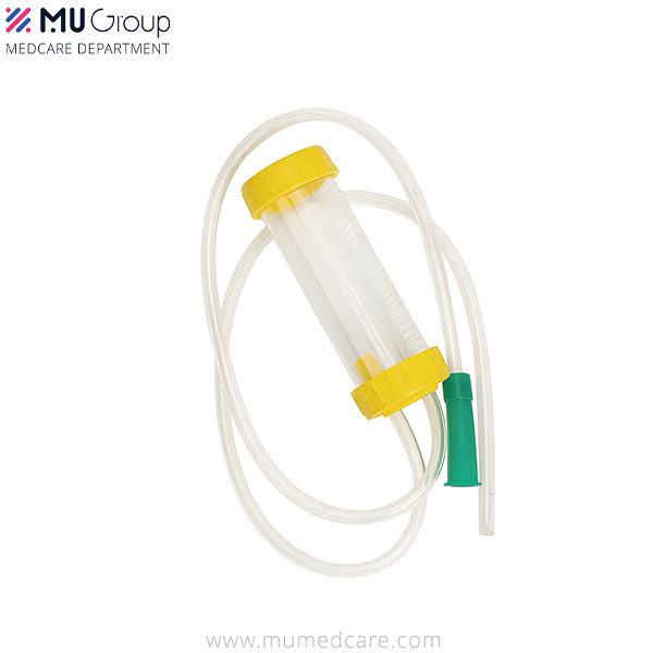
In critical care settings, patient needs frequently overlap. Patients requiring advanced central access often require simultaneous advanced airway management. Standardizing the procurement of related medical supplies creates clinical synergy. For example, a facility upgrading its central line kits should simultaneously evaluate its respiratory inventory, ensuring high-quality items like a closed suction catheter for mechanically ventilated adults or a specialized mucus extractor for pediatric and neonatal airway clearance are sourced from reliable manufacturers. This holistic approach to critical care procurement ensures all life-sustaining interventions are supported by top-tier equipment.
The longevity and safety of any access device depend heavily on post-insertion care. Standardized dressing change kits are essential for maintaining line patency and compliance with infection control protocols. These kits should include antimicrobial patches (such as chlorhexidine-impregnated sponges), sterile transparent dressings, skin protectants, and pre-filled flush syringes. Providing clinicians with all necessary components in a single, standardized package reduces the likelihood of protocol breaches and minimizes the risk of introducing pathogens during routine maintenance. Facilities should audit their supply rooms to ensure these kits are readily available on all acute care units.
Deploying vascular devices carries inherent risks that must be managed through rigorous clinical protocols and continuous education. A proactive approach to complication mitigation protects patients and reduces the financial burden of hospital-acquired conditions. Complications range from minor localized site infections to life-threatening systemic sepsis and massive venous thrombosis.
Preventing CLABSI requires strict adherence to evidence-based insertion and maintenance bundles. This includes utilizing maximal sterile barrier precautions during central line placement (cap, mask, sterile gown, sterile gloves, and a full-body sterile drape). Clinicians must employ chlorhexidine-based skin antisepsis and ensure proper hand hygiene before any manipulation of the line. Furthermore, clinical teams must conduct daily necessity reviews during multidisciplinary rounds, promptly removing any central line that is no longer clinically indicated to minimize exposure time. Scrubbing the hub for at least 15 seconds before every access is a non-negotiable standard of practice.
Catheter occlusion and vessel thrombosis disrupt therapy and can lead to severe systemic complications, including pulmonary embolism. Mitigation strategies include strict adherence to routine mechanical flushing protocols using a push-pause technique to clear the catheter lumen and prevent fibrin buildup. Additionally, utilizing ultrasound guidance during insertion allows clinicians to measure vessel diameter accurately, ensuring an appropriate catheter-to-vein ratio that promotes adequate blood flow around the device, thereby reducing the risk of chemical and mechanical thrombosis.
Institutional success relies on scalable training programs. Establishing dedicated vascular access teams (VATs) significantly improves first-stick success rates, reduces overall complications, and standardizes practices across the facility. Continuous education on ultrasound-guided insertion techniques and advanced troubleshooting ensures that clinical staff remain proficient in handling complex vascular anatomy. Training should include simulated insertions on phantoms before clinicians are permitted to perform procedures on live patients.
Patient involvement is a critical component of access management. Providing clear education regarding the purpose of the device, expected maintenance routines, and signs of potential complications improves patient comfort and compliance. Educating patients on proper outpatient site care, especially for PICCs and tunneled lines, drastically reduces the incidence of accidental dislodgement and community-acquired line infections. Patients should be instructed to keep the dressing dry during showers and to immediately report any redness, swelling, or pain at the insertion site.
No single access device is universally appropriate for all clinical scenarios. Achieving clinical efficacy requires a rigorous evaluation of the infusate characteristics, the projected duration of therapy, and the specific anatomical limitations of the patient. By aligning device capabilities with clinical requirements, healthcare providers minimize complications, preserve venous health, and ensure the uninterrupted delivery of critical therapies. Procurement and clinical committees must prioritize vendors that offer comprehensive product lines engineered for infection reduction, backed by strong clinical education support. Standardizing equipment across departments reduces practice variation and enhances overall patient safety.
Conduct a comprehensive internal audit of current CLABSI rates and PIVC failure rates to identify specific clinical gaps.
Review and update vendor catalogs to ensure access to advanced vascular devices and complementary respiratory equipment.
Implement standardized, ultrasound-guided insertion training for all clinicians responsible for medium and long-term access placement.
Establish daily review protocols in all intensive care units to assess the ongoing necessity of central venous catheters.
A: It is a clinical procedure involving the insertion of a sterile, flexible plastic tube (catheter) into a blood vessel. This allows healthcare providers to draw blood, monitor hemodynamic status, or deliver medications, fluids, and nutrients directly into the patient's bloodstream.
A: Peripheral access involves inserting a short catheter into superficial veins, typically in the hands or arms, for short-term, non-irritating therapies. Central access involves placing a longer catheter that terminates in the large veins near the heart, allowing for the safe delivery of caustic medications, long-term treatments, and rapid fluid resuscitation.
A: A midline catheter is preferred for medium-term therapies (typically up to 29 days) that require reliable access but do not involve continuous vesicants or highly hyperosmolar solutions. Midlines avoid the central circulation, thereby eliminating the risk of central line-associated bloodstream infections (CLABSI) associated with PICCs.
A: Historically, peripheral IVs were replaced every 72 to 96 hours. However, current clinical guidelines often recommend replacing them only when clinically indicated—such as signs of phlebitis, infiltration, or infection—provided the site is rigorously monitored and maintained.
A: IO access is indicated for life-threatening emergencies, such as cardiac arrest, severe shock, or major trauma, when traditional peripheral venous access cannot be established rapidly. It provides immediate, central-equivalent access for fluids and medications but must be removed within 24 hours.
A: Risk factors include prolonged catheter dwell time, insertion in the femoral vein, failure to use maximal sterile barriers during placement, inadequate skin antisepsis, poor hub maintenance, and frequent accessing of the line without proper decontamination protocols.


